
Electron and Neutron Diffraction – 5 captivating difference
Electron and Neutron Diffraction differ in the particles used, wavelength, energy range, sensitivity, sample preparation, experimental setup, and applications. Electron diffraction is particularly useful for studying electronic structures and nanomaterials, while neutron diffraction is valuable for determining atomic positions, magnetic properties, and structures of complex molecules. Both methods can be combined with other techniques for greater comprehension of materials.
Brief overview of Electron and Neutron Diffraction
Electron Diffraction: Electron diffraction, also referred to as Diffraction of Electrons, is a technique which uses electron beams to explore material structures. Diffraction takes advantage of electron’s wave-particle duality which means they act both as particles and waves simultaneously. Electron Diffraction experiments involve using an energetic beam of electrons directed against a sample to analyse its pattern of diffraction for information about both its atomic structure and crystal’s architecture.
Electron diffraction produces patterns due to electron waves interacting with lattice material structures, interfering and diffracting against each other as they pass through it. Electron diffraction has long been used by fields including solid state materials science, physics and nanotechnology for studying crystal structures as well as understanding molecular arrangements within various materials.
Neutron Diffraction: Neutron diffraction is an examination technique using neutron beams to study magnetic and atomic structures within materials. Neutrons, as electrically neutral particles, interact with atomic nuclei to provide valuable insight into where electron density distribution exists in materials. Neutron diffraction can be particularly helpful for studying materials with light elements (such as hydrogen) as well as for characterizing magnetic properties.
Through neutron diffraction, neutron beams are directed at an object and their pattern of diffraction analyzed to discover both their arrangement of atoms as well as magnetization moments. This method has many applications across physics and chemistry as well as materials science and biology for studying crystal structures, magnets and macromolecular assemblies in living systems.
Both techniques offer distinct advantages that complement each other when studying materials and their structures. Electron diffraction provides excellent spatial resolution while being highly sensitive to elements with lighter chemical atoms while neutron diffraction provides information on all atom positions including hydrogen. Together these techniques enable us to better comprehend our microscopic universe while being useful tools in research projects or technological breakthroughs.
Definition of diffraction
Diffraction refers to the bending or spreading of waves as they encounter an obstacle or pass through an aperture. It is a fundamental phenomenon that occurs when waves encounter an obstruction or encounter changes in their medium, resulting in the redistribution of their energy and the formation of patterns. Diffraction can occur with various types of waves, including light waves, sound waves, water waves, and even particle waves like electrons or neutrons.
In the context of electromagnetic waves, such as light, diffraction occurs when the wave encounters an object or an aperture that is comparable in size to the wavelength of the wave. As the wave interacts with the obstacle or aperture, it undergoes interference, resulting in the bending, spreading, and pattern formation.
Diffraction patterns can exhibit a range of characteristics, including the presence of constructive and destructive interference, resulting in regions of increased or decreased wave intensity. These patterns can be observed as alternating bright and dark regions, known as fringes or interference patterns, which can provide valuable information about the nature of the waves, the obstacles encountered, and the size or shape of the diffracting objects.
In scientific research, diffraction is extensively used to study the structure of materials, such as crystals, proteins, and nanoparticles. By analyzing the diffraction patterns produced by waves interacting with these materials, scientists can infer valuable information about their atomic arrangement, molecular structure, and other physical properties.
Diffraction plays an integral part in numerous scientific disciplines, from physics, chemistry, biology and materials science through engineering. Diffraction provides researchers with an efficient means of investigating microscopically-based properties of matter and providing valuable insight into wave behaviour as well as interactions between waves in an environment.
Electron Diffraction
Electron difffraction is a useful technique that utilizes high-energy electron beams to study materials at both molecular and atomic scale. Based on electron’s dual nature of wave and particle properties, electron difffraction provides insight into properties such as both wave-like characteristics as well as particle properties that allow researchers to gain a comprehensive view.
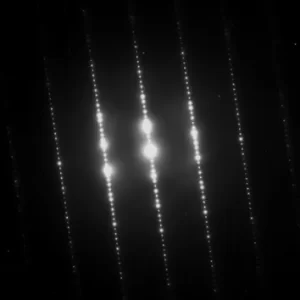
In electron diffraction experiments, a beam of electrons is generated using an electron gun, accelerated to high energies, and focused onto a sample. The electrons interact with the atoms in the sample, and their wave nature causes them to diffract as they pass through or scatter from the atomic structure. This diffraction leads to the formation of an interference pattern, which can be captured and analyzed.
The diffraction pattern obtained in electron diffraction experiments provides valuable information about the arrangement of atoms within the sample. The spacing between atoms and the crystal lattice structure can be determined by analyzing the angles and intensities of the diffracted electron beams. By comparing the observed diffraction pattern with theoretical models, scientists can deduce the atomic arrangement and crystal structure of the material under investigation.
Electron diffraction is widely used in various scientific disciplines. In materials science, it is employed to study the crystal structures of metals, alloys, semiconductors, and other solid-state materials. It can provide insights into the arrangement of atoms, defects, grain boundaries, and phase transformations. Electron diffraction is also utilized in the field of electron microscopy to obtain high-resolution images and diffraction information simultaneously, enabling the study of nanostructures and individual nanoparticles.
Additionally, electron diffraction has applications in the study of molecules and biological structures. It is used to determine the structures of organic compounds, proteins, and complex biomolecules. By analyzing the electron diffraction patterns, scientists can derive important structural information, such as bond lengths, angles, and overall molecular shapes.
Electron diffraction is a versatile technique that allows scientists to explore the atomic and molecular structure of materials. It has played a significant role in advancing our understanding of the microscopic world and has applications in various scientific fields, ranging from materials science and solid-state physics to chemistry and structural biology.
Geometrical considerations of Electron Diffraction
Geometrical considerations play a crucial role in electron diffraction experiments and the interpretation of diffraction patterns.
Here are some key geometrical considerations in electron diffraction:
- Bragg’s Law: Bragg’s Law is a fundamental principle in diffraction that relates the angle of incidence, the angle of diffraction, and the spacing between crystal planes in a material. It is expressed as nλ = 2dsinθ, where n is the order of the diffraction, λ is the wavelength of the electrons, d is the spacing between crystal planes, and θ is the diffraction angle. This law determines the positions and angles at which diffracted electron beams are observed in the diffraction pattern.
- Crystal Orientation: The orientation of the crystal lattice with respect to the incident electron beam is critical in electron diffraction. Different crystal orientations result in different diffraction patterns. By rotating the crystal and recording diffraction patterns at various angles, it is possible to obtain a complete understanding of the crystal’s three-dimensional structure.
- Zone Axis: The zone axis refers to the direction in which the crystal planes are parallel to the incident electron beam. When the crystal is oriented such that the electron beam is along a high-symmetry direction, it can produce strong and well-defined diffraction spots in the pattern. Determining the zone axis is essential for obtaining clear and interpretable diffraction patterns.
- Sample Thickness: The thickness of the sample plays a role in electron diffraction experiments. Thin samples allow for better visibility of the diffracted beams and minimize the effects of multiple scattering and dynamical scattering. However, thicker samples can still produce diffraction patterns, albeit with more complex and less resolved features.
- Aperture Size: The size of the aperture or detector used to capture the diffraction pattern affects the resolution and intensity of the diffraction spots. Smaller apertures can provide higher angular resolution but may reduce the intensity of the diffracted beams. Larger apertures allow for better intensity but may result in broader diffraction spots.
- Camera Length: The camera length, which is the distance between the sample and the detector, affects the magnification and scale of the diffraction pattern. Adjusting the camera length allows for optimization of the diffraction pattern’s size and resolution, ensuring that all relevant information is captured.
By carefully considering these geometric factors, scientists can accurately interpret electron diffraction patterns and extract valuable structural information about the crystal lattice, including unit cell dimensions, symmetry, and atomic arrangements.
Neutron Diffraction
Neutron diffraction is an extremely effective research technique which utilizes neutron beams for studying the atomic and magnetic structures of various materials. As they do not conduct electricity, neutrons penetrate deep within structures to gather more information on where electron concentration exists in their structure.
Neutron Diffraction Testing involves creating an ion beam using either spallation or nuclear reactor sources and sending it towards an important object, where its nuclei collide with it and cause scattering that produces an interesting diffraction pattern that can be observed and studied.
Neutron diffraction tests reveal details regarding the structure of molecules within a sample by studying its patterns of diffraction. By analyzing angles and intensities of diffracted neutron beams, scientists can pinpoint precise locations within structures containing lighter elements like hydrogen. Diffraction is particularly advantageous when studying materials that include lightweight elements which cannot easily be studied with traditional means such as electron microprobe analysis or physical testing methods.
One of the hallmarks of neutron difffraction is its sensitivity towards magnetic properties. Neutrons themselves have magnetic fields and this interaction with samples’ magnetite momenta can be observed through its diffraction pattern allowing researchers to investigate properties like ordering, domain structures and phase changes within materials studied through neutron difffraction techniques.
Neutron diffraction can be utilized across many scientific fields. Solid state material science and physical physics utilize neutron diffraction for studying crystal structures, phase shifts and magnetism behaviour; whilst in chemistry neutron difffraction provides insights into molecular structures as well as chemical bonds; finally it also assists researchers studying macromolecules such as nucleic acids or proteins to explore three-dimensional structures and understand roles more fully.
Neutron diffraction is a versatile technique that allows scientists to probe the atomic and magnetic structures of materials. Its ability to penetrate deeply into samples and its sensitivity to light elements and magnetic properties make it a valuable tool in various scientific fields, contributing to our understanding of the microscopic world and driving advancements in materials science, chemistry, and biology.
Key Differences Between Electron and Neutron Diffraction
Electron diffraction and neutron diffraction are both powerful techniques for studying the atomic and molecular structures of materials, but they have several key differences.
Here are some of the main differences between electron diffraction and neutron diffraction:
- Nature of the particles used:
- Electron diffraction involves using a beam of high-energy electrons.
- Neutron diffraction utilizes a beam of neutrons, which are electrically neutral particles found in atomic nuclei.
- Wavelength and energy differences:
- Electrons have much shorter wavelengths than neutrons. De Broglie electrons’ wavelength typically measures in picometers while their de Broglie spectrum lies on an order of angstroms scale.
- Electron energies used for diffraction experiments tend to lie between several tens and hundreds of Kiloelectron Volts (keV), while neutrons can produce energies between thermal (meV) and many hundreds of Kiloelectronvolts (keV).
- Sensitivity to different types of atomic interactions:
- Electron diffraction is sensitive to the distribution of electron density within a material and is useful for studying light elements and their electronic structures.
- Neutron diffraction is sensitive to the positions of atomic nuclei and the magnetic properties of materials. It provides information about the positions of all atoms, including light elements like hydrogen, and is particularly useful for studying magnetic materials.
- Sample preparation requirements:
- Electron diffraction typically requires samples to be prepared as thin foils or powders that can withstand the high vacuum environment of the electron microscope or diffraction apparatus.
- Neutron diffraction can work with a broader range of sample types, including bulk materials, liquids, and even some biological samples. However, samples often need to be large enough to interact significantly with the neutron beam.
- Experimental considerations and limitations:
- Electron diffraction experiments are typically performed in high-vacuum environments and require sophisticated electron optics, such as electron microscopes or specialized diffractometers.
- Neutron diffraction experiments often take place at large-scale research facilities, such as nuclear reactors or spallation sources, due to the need for a high-intensity neutron source. These facilities are not as widely available as electron microscopy facilities.
It’s worth noting that despite these differences, both electron diffraction and neutron diffraction techniques play crucial roles in studying the structures of materials and have complementary strengths. They provide valuable insights into different aspects of atomic and molecular arrangements, making them indispensable tools in various scientific disciplines.
Comparison chart between Electron and Neutron Diffraction
| Aspect | Electron Diffraction | Neutron Diffraction |
|---|---|---|
| Particle Used | Electrons | Neutrons |
| Wavelength | Short (picometers) | Longer (angstroms) |
| Energy Range | Tens to hundreds of keV | Thermal (meV) to tens of keV |
| Sensitivity | Electron density | Atomic positions, magnetic properties |
| Sample Types | Thin foils, powders | Bulk materials, liquids, some biological samples |
| Experimental Setup | Electron microscope, specialized diffractometers | Large-scale facilities with nuclear reactors or spallation sources |
| Application Range | Crystal structures, nanomaterials, electron microscopy | Crystal structures, magnetic materials, chemical bonding, biomolecules |
| Sample Preparation | High-vacuum environment, thin samples | Wide range of sample types, large enough to interact significantly with neutrons |
| Availability | Electron microscopy facilities widely available | Neutron sources limited to large-scale research facilities |
| Complementary Nature | Can be used in combination with other techniques | Can be used in combination with other techniques |
Similarities Between Electron and Neutron Diffraction
While electron diffraction and neutron diffraction have several key differences, there are also some similarities in their principles and applications.
Here are some of the similarities between electron diffraction and neutron diffraction:
- Principle of diffraction: Both methods utilize the concept of diffraction. Electrons and neutrons interact with material atoms through waves (which scatter off of it and lead to destructive and constructive interference patterns), creating patterns known as diffraction.
- Analysis of interference patterns: In both electron diffraction and neutron diffraction, the resulting diffraction patterns are analyzed to extract information about the atomic and molecular structure of the material. The angles and intensities of the diffracted beams are measured and used to determine the arrangement of atoms, crystal symmetry, and other structural parameters.
- Determining atomic structures: Both techniques are powerful tools for determining the atomic structures of materials. They can provide valuable information about the positions of atoms within a crystal lattice, the presence of defects or impurities, and the bonding between atoms.
- Crystallography applications: Electron diffraction and neutron diffraction are extensively used in crystallography to study the structures of crystalline materials. They can be used to determine the unit cell dimensions, crystal symmetry, and atomic coordinates of crystal structures.
- Applications in material science: Both approaches may be applied in materials science for analyzing various kinds of materials such as alloys, metals, ceramics, semiconductors and polymers. By understanding relationships among physical properties, atomic structure and material behavior they help provide insight into material behavior.
- Complementary techniques: Electron and neutron difffraction techniques are frequently combined with other analytical methods, like X-ray diffraction and spectroscopy, in order to gain greater insight into material properties. By employing multiple diffraction techniques at once, one can get a comprehensive view of structural materials’ characteristics.
Although electron diffraction and neutron diffraction differ in the nature of particles used and their specific strengths, they share common ground in their principles of diffraction and their application in the investigation of atomic and molecular structures. Their complementary nature makes them valuable tools in the field of structural characterization and materials research.
Conclusion
Diffraction of electrons and neutrons provides effective analysis of molecular and atomic structures of various materials, from those with different molecular or atomic composition to complex compounds with complex interfacial interactions. Electron diffraction uses high energy electron beams while neutron difffraction relies on neutron beams; both methods differ due to particle type employed, wavelength/energy characteristics as well as sensitivities to various interactions, sample preparation needs and any special considerations related to experiments.