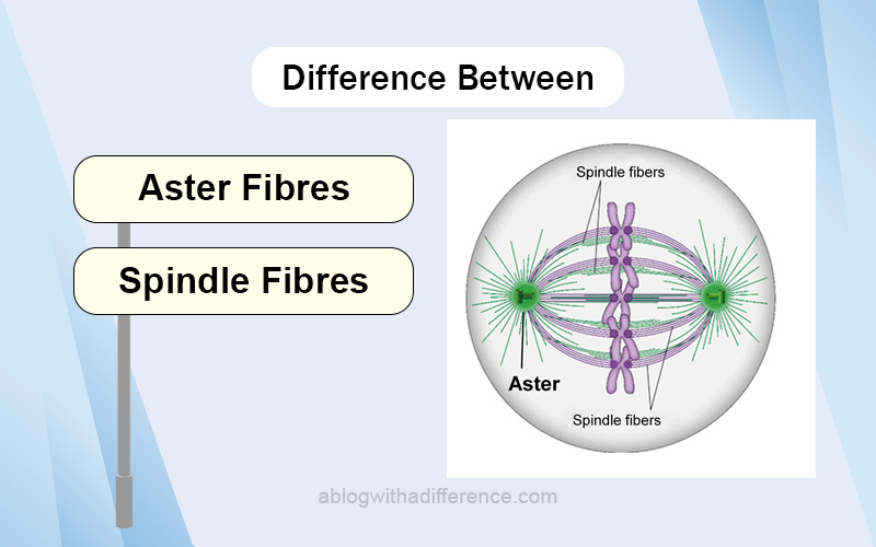
Difference Between Aster and Spindle Fibres
Aster and Spindle Fibres are vital in cell divisions, meiosis, and mitosis. Both of them are part of the spindle apparatus. The Spindle apparatus comprises spindle fibers, motor protein, and chromosomes. Animal cells comprise microtubule arrays. Spindle fibers and Aster fibers are microtubules.
A brief overview of cell division and its importance
Cell division is an essential process that enables organisms to adapt, thrive, and repair damaged tissues, essential for life and maintaining continuity. Cell division takes place when one parent cell divides into two or more daughter cells each with complete genetic information inherited faithfully by subsequent generations of cells.
Cell division plays an essential part in many biological processes, including:
Cell division plays an essential role in both the growth and development of organisms as they expand in size and form new tissues and organs. cellule And when tissues have been injured due to injury or disease, the division helps replace damaged cells to speed healing by replacing these with healthy ones and speeding recovery time.
Cell division is often the main method of reproduction among single-celled organisms and certain multicellular organisms, providing them with the means of procreating and expanding their populations.
In multicellular organisms, cell division enables constant renewal of tissues and organs by way of cell division; this ensures proper function is sustained across their lifespans.
Cell division in sexual reproduction contributes significantly to genetic diversity by shifting and combining genes in meiosis.
Understanding cell division mechanisms and regulation is integral for studying developmental biology, cancer research, regenerative medicine, and various other fields of biology and medicine. Furthermore, abnormalities in cell division can result in genetic disorders, developmental abnormalities, or diseases like cancer; thus understanding all its complexities plays a pivotal role in furthering knowledge of life processes as well as meeting various health-related challenges.
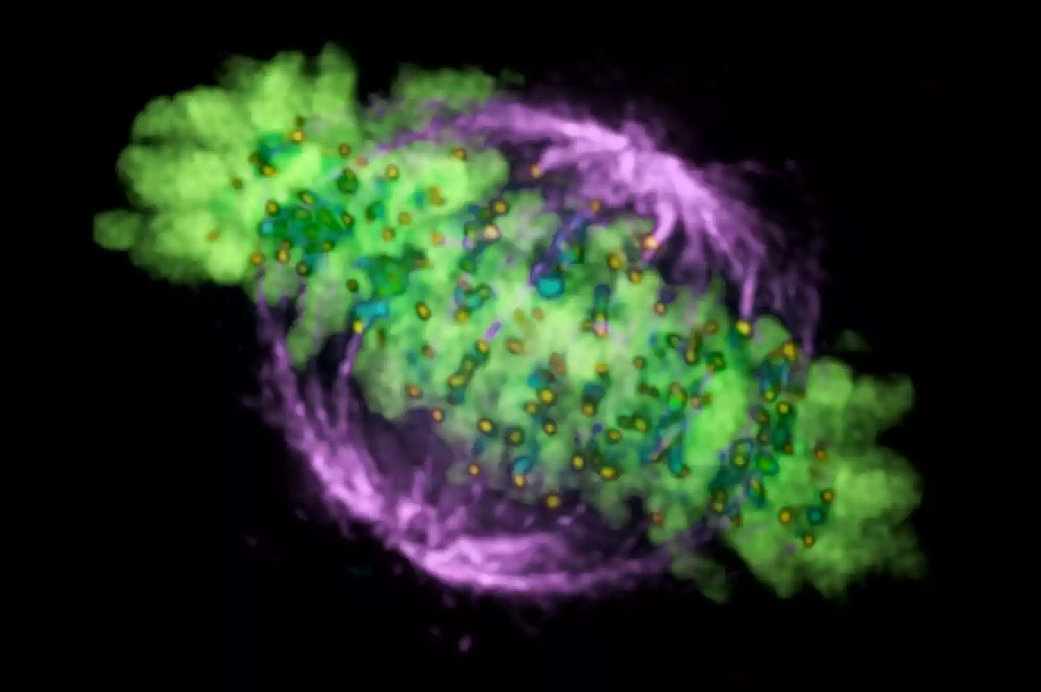
What are Aster Fibres?
Aster fibers are cellular star-shaped structures comprised of a centrosome. They’re connected to microtubules in the early meiotic and mitotic stages within animal cells. Aster fibers are composed of microtubules and form a component of the cell cytoskeleton.
Aster fibers do not form in plant cells. Microtubules compose the Aster Rays and emanate from the centrosome. They provide support for the chromosomes, guiding them into the correct place in cell division.
They assist the chromosomes in getting get arranged and align at the center of the nucleus. Aster fibers transfer half the genetic information on both sides of the nucleus in the process. Consequently, the nucleus is divided into two halves, leaving two nuclei that are daughters.
The first appearance of aster fibers is during the cell division process called prophase. Asters in two centrosomes move to opposite sides of the nucleus, forming mitotic spindles. The aster fibers form the spindle fibers that extend out across both poles as well as the fibers which attach to chromosomes via the Kinetochores.
The nuclear envelope splits into fragments and mitotic spindles are formed during metaphase. Spindle fibers transport the chromosomes toward the center during metaphase. The direction of the aster fibers determines the direction of the division of cells throughout cell division.
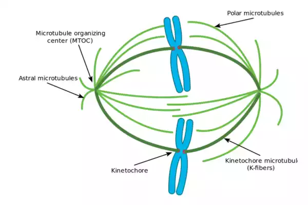
Astral microtubules
Astral microtubules comprise an unrelated microtubule type they only form at the time of mitosis and shortly before. They can be described as any microtubule that originates from the centrosome but is not connected to the Kinetochore.
Astral microtubules form within the actin skeleton, and connect to cells’ cortex, helping in the orientation of spindles. They form circular arrays surrounding the centrosomes. The rate of turnover for this microtubule family is much higher than the other populations.
The function of astral microtubules can be assisted by specific dyneins to the function. They have lightweight chains (static parts) that are attached to cell membranes, as well as their globular components (dynamic parts) connected to microtubules.
The chains in the globular form attempt to go toward the centrosome. However, since they are connected to the cell membrane they pull the centrosomes toward the membrane and thereby aiding the process of cytokinesis.
Astral microtubules do not have to be present in the development of mitosis. They must be present to guarantee the integrity of the process. Astral microtubules’ function is generally regarded as the determination of the geometry of cells.
They are essential to ensure proper positioning and orientation to the mitotic spindle They are also essential in the determination of cell division location based on the shape and polarity of cells.
The existence of astral microtubules is dependent upon the centrosomal integrity. It is also dependent on several microtubule-associated proteins such as EB1 and adenomatous polyposis coli (APC).
What are Spindle Fibres?
Spindle fibers are microscopic tubules that create the meiotic and mitotic spindle in cell division. These are protein-based structures and also they split the genes in cells. Spindle fibers can also be referred to as mitotic spindles in mitosis because they create identical genetically identical daughter cells as well as meiotic spindles during meiosis.
After all, it creates gametes having only half of the number of chromosomes as the parent cell. Thus, spindle fibers are able the ability to divide their chromosomes equally into two cells that are derived from a parent division of cells.
In this manner, Spindle fibers can move their chromosomes through attachment to centromeres and chromosomal arms. When in prophase, spindle fibers develop at the opposing cells’ poles as cells expand, spindle fibers expand. Sister chromatids join spindle fibers through their kinetochores.
When metaphase is occurring, spindle fibers are known as polar fibers which extend outwards from the cell’s poles to the middle point. Chromosomes are placed at the center by the pull of spindle fibers. When anaphase occurs, spindle fibers reduce in size, while sister chromatids pull towards opposite poles. In telophase, spindle fibers are dispersed as chromosomes split.
Spindle structure
The attachment of microtubules to the chromosomes happens through the kinetochores who actively keep track of spindle development and stop early anaphase start. Microtubule polymerization and depolymerization are dynamic processes that cause chromosome congression.
Depolymerization of microtubules causes tension at kinetochores. [33. bipolar attachment of sister kinetochores microtubules that originate from opposite cell poles combine opposing tension forces, thereby aligning the chromosomes near the cell’s equator before allowing them to segregate into daughter cells. After every chromosome has been bi-oriented, anaphase begins with the onset of anaphase. cohesin that binds sisters chromatids is broken and allows the movement of twin chromatids towards opposite poles.
The cellular spindle apparatus comprises the spindle microtubules as well as related proteins, such as dynein and kinesin molecular motors condensed chromosomes and any asters or centrosomes which may be found at the poles of the spindle, based according to the cell type.
[4The spindle apparatus is a vaguely ellipsoid cross-section and taper towards the end. In the broad middle part called the midzone of the spindle, the antiparallel microtubules have been bundled by Kinesins. At the ends with pointed ends, also known as the spindle poles, microtubules get formed by centrosomes found in the majority of animals’ cells.
The Acentrosomal (also known as astral spindles do not have centrosomes or asters in the spindle poles or asters, respectively, and are found for example, during meiosis in the majority of species of animals. [5] In this case, the GTP gradient of Ran is the primary control for spindle microtubules’ organization and assembly. When it comes to fungi the spindles develop within spindle poles which are encased in the nuclear envelope and do not disintegrate during mitosis.
Differences Between Aster and Spindle Fibers
A summary of the differences between aster and spindle fibers can be provided as follows:
1. Location and Distribution:
Aster fibers: These filamentous structures, characteristic of animal cells, radiate outward from centrosomes near their nuclei towards cell peripheries. They encase centrosomes located nearby.
Spindle fibers: Spindle fibers form part of the spindle apparatus, an essential structure in cell division. Their length spans from pole to pole within cells while connecting centrosomes and chromosomes throughout them all.
Function and Purpose of Aster Fibers: Aster fibers play an integral part in positioning the spindle apparatus during cell division, helping ensure accurate chromosome segregation and precise spindle orientation for accurate cell division.
Spindle Fibers: Spindle fibers play a pivotal role in cell division by aiding with the movement and segregation of chromosomes during cell division. Attaching themselves to chromosomal kinetochores, spindle fibers exert forces that move chromosomes to opposite poles of their host cell and force segregation of DNA from them towards opposite sides. Their structure and composition vary among species but the most prevalent examples include silkworm cells (Drosophila melanogamy).
2. Function and Purpose:
Aster fibers: Aster fibers consist of polar microtubules extending from centrosomes. Like any microtubule, Aster fibers contain tubulin protein subunits arranged into hollow cylindrical structures for easy transport through tissue layers.
Spindle Fibers: Spindle fibers are composed of tubulin protein subunits organized as microtubules. There are two major categories of these microtubules; these are known as Kinetochore fibers which connect chromosomes directly with spindle poles; while Polar fibers overlap centrally throughout a spindle and form its central region.
3. Interaction with Other Cellular Components:
Structure and Composition:
Aster fibers: These filamentous proteins interact with various cell components, including its cortex and cytoplasmic proteins, to assist with the positioning of the spindle apparatus and ensure its proper organization.
Spindle fibers: Spindle fibers interact directly with chromosomes at their protein structures called kinetochores, exerting forces that enable their movement and segregation. Their significance: Spindle fibers exert forces upon chromosomes which facilitate segregation processes. Throughout development, they exert forces that facilitate cell division. tiennent WHY: They play such an essential role.
Comparison chart
Here is a comparison chart of differences between aster fibers and spindle fibers:
| Topic | Aster Fibers | Spindle Fibers |
|---|---|---|
| Location | Surround the centrosomes in animal cells | Found within the spindle apparatus during cell division |
| Function | Organize and position the spindle apparatus | Facilitate movement and segregation of chromosomes |
| Structure | Composed of polar microtubules | Composed of microtubules (kinetochore and polar fibers) |
| Interaction | Interact with cell cortex and cytoplasmic proteins | Interact with chromosomes at kinetochores |
| Importance | Contribute to proper spindle orientation | Ensure accurate chromosome segregation |
| Distribution | Radiate from centrosomes towards the cell periphery | The span from pole to pole within the cell |
| Examples | Prominent in animal cells during mitosis | Present in various organisms undergoing cell division |
Similarities between Aster and Spindle Fibres
Aster fibers and spindle fibers are two microtubule structures involved in cell division.
While their characteristics and functions differ considerably, both share some key similarities:
- Composed of microtubules: Both aster and spindle fibers contain microtubules – tubular structures composed of protein subunits known as tubulin that make up their constituent parts.
- Dynamic Behavior: Both fiber types exhibit dynamic instability, meaning their growth and shrinkage is continuously fluctuating over time. This dynamic behavior allows these cells to explore cellular space while making connections and exerting force during cell division.
- Tubulin Protein Subunits: Both aster and spindle fibers consist of tubulin protein subunits arranged in an open cylindrical pattern characteristic of microtubules.
- Engaged in cell division: Aster fibers and spindle fibers play an integral part in cell division processes, assuring accurate chromosome segregation and successfully concluding cell division processes.
- Interaction With Cell Components: Both types of fibers interact with different cellular components to fulfill their functions, with Aster fibers engaging the cell cortex and cytoplasmic proteins to position the spindle apparatus, while Spindle fibers directly interact with chromosomes at specific structures called kinetochores.
Aster fibers serve a primary function in organizing and positioning of the spindle apparatus while spindle fibers promote the segregation of chromosomes during cell division processes, yet both share similar microtubule-based structures to ensure smooth distribution and division processes.
Conclusion
Aster and spindle fibers are two forms of microtubule structures involved in cell division. Animal cells contain aster fibers encasing centrosomes; this essential structure is necessary to organize and position the spindle apparatus properly for cell division. Spindle fibers play an integral part in maintaining the proper orientation of the spindle apparatus and ensuring accurate chromosome segregation during cell division.
By contrast, spindle components contribute to proper orientation by contributing their proper positions within it – aiding with the movement of chromosomes from one cell division cycle to the next. Aster fibers attach to chromosome kinetochores and exert forces that pull chromosomes toward opposite poles of a cell.
Spindle fibers consist of two kinds of threads – kinetochore fibers connecting chromosomes with spindle poles, as well as polar threads spanning across its center region – connecting aster fibers with various aspects of cell division and segregation processes, providing insight into cell biology processes as well as diseases in general. Understanding these differences will bring greater insight into the complex processes involved.